
Patient guide · Understanding your MRI
Your MRI report, in plain English. Almost every scary word has a boring meaning.
Radiology reports are written by one doctor for another, in a shorthand built to be precise, not comforting. This page translates it: what the words mean, which ones matter, and which ones sound far worse than they are.
L4-L5: Disc desiccation with loss of disc height. Broad-based posterior disc protrusion with a superimposed left paracentral extrusion measuring 7 mm, effacing the thecal sac and impinging the traversing left L5 nerve root. Mild facet arthropathy. Mild bilateral foraminal narrowing.
L5-S1: Annular fissure with high-intensity zone. Modic endplate changes.
IMPRESSIONDegenerative changes, most pronounced at L4-L5. No acute findings. Clinical correlation recommended.
Tap any highlighted word to jump to its meaning. A sample written for this page, not a real patient's report.
Why this page exists
Nobody ever showed you the pictures. There's a reason.
The doctor most trained to read your MRI, the radiologist, writes the report and never meets you. The doctors who do meet you usually work from that report, and a surgeon's schedule rarely has room to sit down and walk through the images with you. Nobody did anything wrong, and you still ended up owning a spine you've never seen.
We think that's backwards.
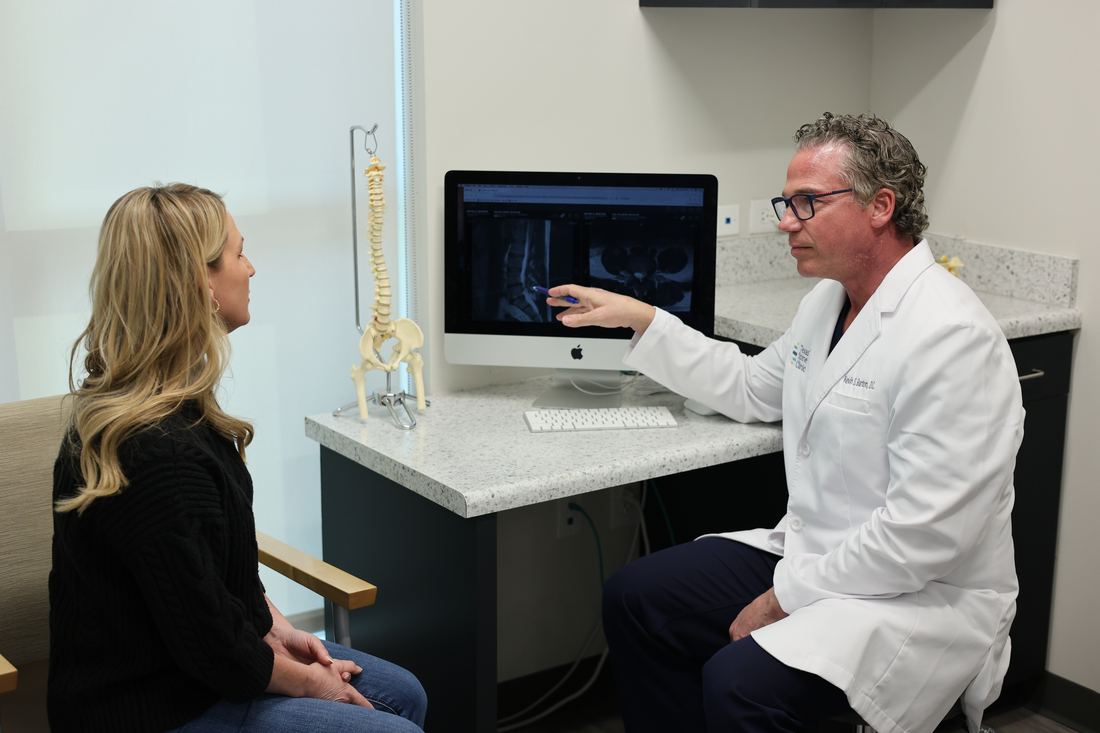
At Texas Spine Clinic your consultation happens in front of a screen with your MRI on it. We go through the pictures, not just the report, and show you what's pressing on what, why it hurts the way it does, and what we plan to do about it.
Start here
First, the disc itself
Between each pair of bones in your spine sits a disc, a shock absorber. Picture a jelly donut with a very tough shell: a ring of hard fibers called the annulus, around a soft, water-rich gel called the nucleus. A healthy disc is roughly 90% fluid. That water is what lets it absorb load.
Almost every word in your report describes one of three things:
How much water is left
Words like desiccation and loss of disc height. A drier disc is a flatter disc.
Whether the shell gave way
Words like bulge, protrusion, extrusion and annular fissure. How far the gel has pushed out, and through what.
What it's touching now
Words like impinging, thecal sac and foraminal. This is the part that decides whether it hurts.
How far it came out
Four words, one ladder
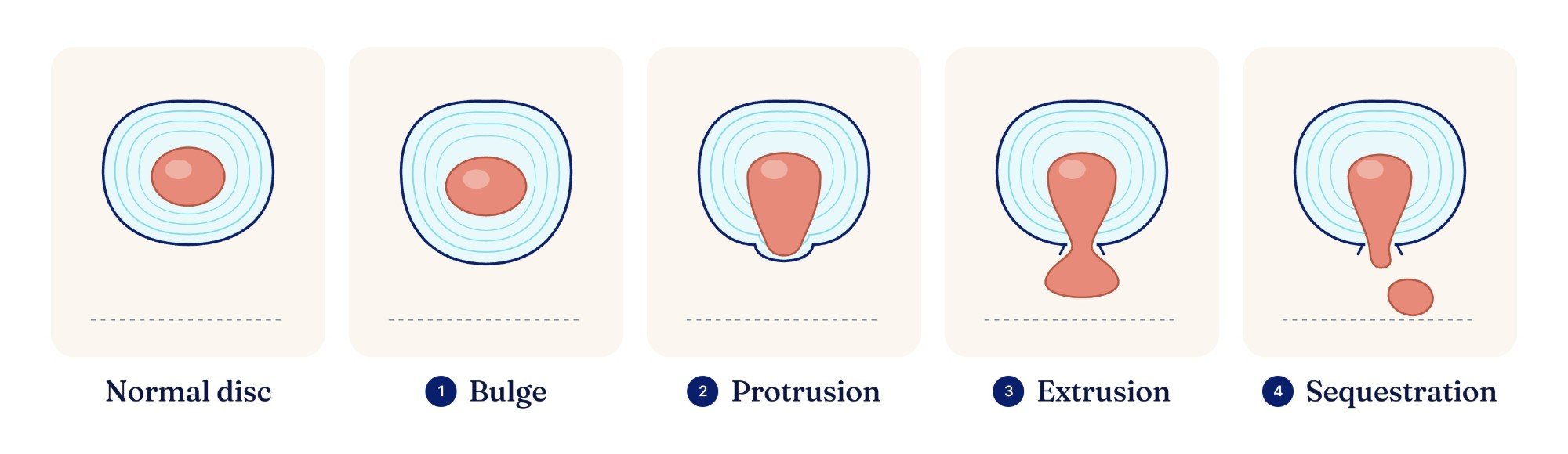
Bulge, protrusion, extrusion, sequestration. These aren't four different diseases. They describe increasing amounts of the same thing.
1Bulge
The whole disc spreads past its normal edge, like a tire low on air. The shell is intact. A bulge is not a herniation, and most never become one.
Bulging discs →2Protrusion
Gel pushes into part of the shell and sticks out, but the base is still wider than the lump. Like pressing your thumb into the inside of a balloon.
3Extrusion
The material squeezes all the way through, and the part outside is wider than the neck it came through. Toothpaste mushrooming out of a small hole.
Herniated discs →4Sequestration
Also called a "free fragment." A piece has broken off completely and sits loose in the canal, no longer attached to the disc.
Bigger on paper isn't more painful.
"Extrusion" sounds much worse than "protrusion," and it is bigger. It is not automatically more painful.
The dramatic ones often shrink.
Large herniations are often the ones most likely to shrink over time. Will it heal on its own?
A fragment that has already broken free is a different conversation. Depending on your symptoms and the fragment's size, it can be a reason we won't treat, which is one more reason we look at your actual imaging before anyone talks about a plan.
The part nobody tells you
The millimetres matter less than where it's pointing
Your report may say 3mm, 5mm, 8mm, 11mm. That's how far the disc material extends past where the disc edge should be. Bigger numbers mean more crowding.
But the number is less important than the direction. We have seen both of these, many times:
Measurements are estimates read off a single slice. Two radiologists can read the same scan a millimetre or two apart, so "6mm" becoming "7mm" on a different machine a year later doesn't mean you're getting worse.
Central
Straight back into the middle of the canal
There's the most room here, so central herniations are sometimes surprisingly quiet, or cause back pain and stiffness without much leg pain.
A small herniation in a tight place beats a big herniation in a roomy place, every time.
Size is the headline. Location is the story.
Check your report against your body
The levels, and why they should match your symptoms
Levels are written as two bones with a dash. The two most common herniations in the body are L4-L5 and L5-S1, the bottom two levels of the lower back, which carry the most load.
- Where you feel it
- Front of the thigh, across the knee, down the inside of the shin.
- Weakness shows as
- A knee that gives out.
- Where you feel it
- Outside of the calf, across the top of the foot, into the big toe.
- Weakness shows as
- Trouble lifting the front of your foot ("foot drop").
- Where you feel it
- Back of the thigh and calf, the heel, the outer foot and little toes.
- Weakness shows as
- Trouble pushing off, or standing on your toes.
- Where you feel it
- C6: down the arm into the thumb and index finger. C7: into the middle finger.
- Read more
- Neck pain and arm symptoms
Word finder
Find the word in your report
Type it in, or filter by type. Each one opens to the plain-English version.
The discBulgeThe whole disc spreads a little past its edge. Shell intact.
Like a tire low on air. A bulge is not a herniation, and most never become one. Bulging discs.
The discProtrusionGel pushing out, with a base wider than the lump.
A broad, contained lump, like a thumb pressed into the inside of a balloon. Step 2 of the ladder.
The discExtrusionMaterial squeezed through the shell and mushroomed out.
The part outside is wider than the neck it came through. Bigger than a protrusion, but not automatically more painful, and large ones are often the most likely to shrink. Step 3 of the ladder.
The discSequestration / free fragmentA piece has broken off and sits loose in the canal.
No longer attached to the disc. This one changes the conversation about treatment, which is why we look at the imaging before anyone talks about a plan.
The discBroad-based vs. focalHow wide a stretch of the disc edge is pushing out.
"Focal" means a narrow section, under a quarter of the disc's edge. "Broad-based" means a wider section, a quarter to half of it. It describes shape, not severity.
The discDesiccationThe disc has lost water. That's all.
Dried out. It shows up darker on the scan. Extremely common, including in people with no pain.
The discLoss of disc height / disc space narrowingThe disc is flatter, so the bones sit closer together.
Usually follows desiccation: less water, less height. Degenerative disc disease.
The discAnnular fissure / annular tearA crack in the disc's tough outer shell.
"Tear" makes people picture a ripped muscle. It's closer to a crack in a rubber gasket. Many people have them without pain, but they can be a source of deep, hard-to-pin-down back pain.
The discHigh-intensity zone (HIZ)A bright spot on the scan, usually marking a fissure.
It's the way an annular fissure often shows up on certain MRI images.
The discSchmorl's nodeA small dent where disc material pushed into the bone.
Usually old, usually silent, frequently reported and almost never the problem.
DirectionCentralStraight back into the middle of the canal, where there's the most room.
Sometimes surprisingly quiet. See it on the diagram.
DirectionParacentral / posterolateralBack and slightly to one side, right where a nerve root sits.
The most common direction by far, because the ligament down the middle of the spine is strongest in the center and pushes material sideways. The classic sciatica herniation. See it on the diagram.
DirectionForaminalInto the small side tunnel where the nerve leaves the spine.
A tight space with nothing to spare. Small foraminal herniations cause outsized pain, and they're the ones most often underestimated from the number alone.
DirectionFar lateral / extraforaminalOutside the side tunnel entirely.
Less common, and it pinches a different nerve than you'd expect from the level, which is why some people's symptoms don't seem to "match" their scan until someone explains this.
Nerves & spaceContacting / abutting / impinging / displacingDegrees of the same thing: touching, crowding, pressing, shoving.
"Impinging" means the disc is pressing on a nerve. That's the electric pain, the burning, the tingling, the numb toes.
Nerves & spaceTraversing vs. exiting nerve rootWhich of two nearby nerves the disc is crowding.
At each level, one nerve is passing by on its way down (traversing) and one is leaving through the side tunnel (exiting). At L4-L5, a paracentral herniation usually crowds the traversing L5 root; a foraminal one crowds the exiting L4 root. That's why direction changes which part of your leg hurts.
Nerves & spaceThecal sac / effacementThe sleeve around the nerve bundle is being indented.
The thecal sac is the fluid-filled sleeve that holds the nerves. "Effacement of the thecal sac" means it's being flattened from one side. It sounds violent. It means indented.
Nerves & spaceForaminal narrowing / stenosis"Stenosis" just means narrowing.
Canal stenosis: the central tunnel is tighter. Foraminal stenosis: the side exit is tighter. Spinal stenosis.
Nerves & spaceLigamentum flavum thickeningA ligament inside the canal has thickened and takes up room.
Space that used to belong to the nerves. Often part of the stenosis picture.
Bone & jointsModic changes / endplate changesChanges in the bone just above and below a disc. Common. Not cancer.
They show where the bone has been taking extra load next to the disc.
Bone & jointsFacet arthropathy / facet hypertrophyArthritis in the small joints at the back of the spine.
The joints are irritated or have thickened up. Very common alongside a disc that has lost height. Facet syndrome.
Bone & jointsSpondylolisthesisOne vertebra has slipped forward on the one below.
Graded 1 through 4. This one changes the plan, and it's something we screen for. What matters isn't the grade but whether the slip is stable, which we check with flexion-and-extension X-rays.
Bone & jointsSpondylosisA general word for wear-and-tear changes.
Not a diagnosis so much as a description.
Report phrasesDegenerative changesMileage, not a prophecy.
The most over-frightening phrase in radiology. Here's what it actually means.
Report phrasesMild / moderate / severeHow much a space has narrowed, not how much you hurt.
These grade the picture. Plenty of people with "severe" on paper feel fine, and plenty with "mild" are in real pain. Your exam decides what the grade means for you.
Report phrases"No acute findings"Nothing broken, nothing that needs an operating room today.
It is not the same as "nothing is wrong with you," and it is not a verdict on your pain.
Report phrases"Clinical correlation recommended""Someone should check this against an actual exam."
The radiologist is saying the picture alone can't tell the whole story. We agree, and it's the reason every consultation starts with an examination.
No match. Try a shorter piece of the word, or bring the report in and we'll go through it with you.
The phrase that scares people most
"Degenerative changes" describes mileage, not a prophecy
If you're in your twenties, thirties or forties, this is probably the line that scared you most. Somebody read "degenerative disc disease" and pictured a spine crumbling on a schedule.
Here's what it actually describes. A radiologist compares your spine to a young, textbook-perfect one, and anything less gets called degenerative. Your disc has lost some water and some height under years of load. That's the whole meaning.
It is extraordinarily common, and it starts early. Disc drying shows up on the scans of plenty of people in their twenties and thirties who feel perfectly fine.
It does not march forward on a timetable. What drives it is load, and that's the part you can change. Degenerative disc disease, fully explained.
Your imaging still matters: bulges, protrusions, extrusions and degeneration are more common in people who hurt.1 A finding just isn't, on its own, proof of what's causing your pain.
Found on the MRIs of people with no back pain at all
33 imaging studies, 3,110 people with zero back pain.2
Disc degeneration
Disc bulges
The scariest words in your report are also printed on the reports of people out playing golf right now.
He's sat on both sides of the screen
What a real MRI review looks like
In February 2025, Dr. Barton's own MRI showed a 15 mm L4-L5 extrusion, one of the worst he had seen in over 20 years of practice. He treated it on the DRX9000. By August, the same level looked like the picture on the right.
That's the kind of conversation your consultation is: your images on the screen, the arrows where they belong, and a plan you understand.
Watch his series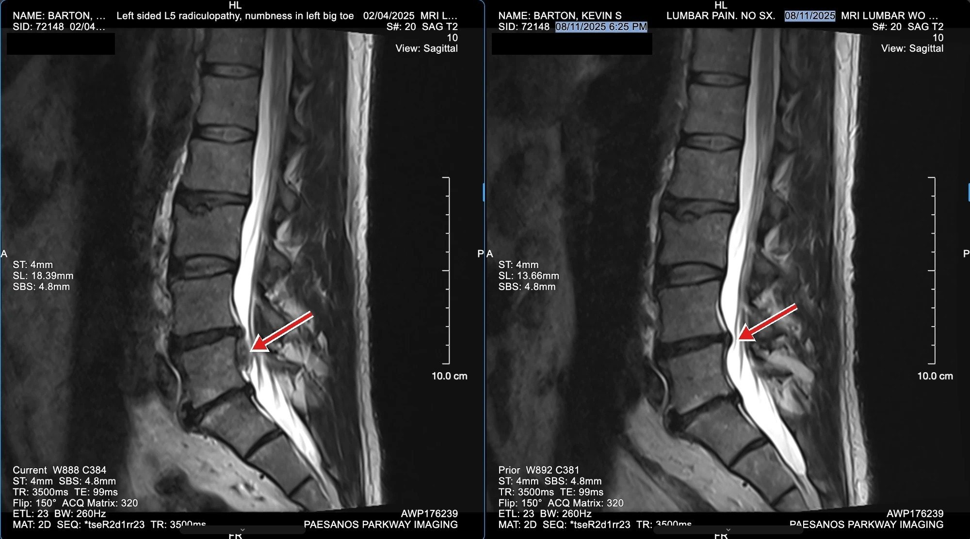
Sources
- Brinjikji W et al. MRI findings of disc degeneration are more prevalent in adults with low back pain than in asymptomatic controls. AJNR Am J Neuroradiol 2015;36(12):2394–2399. ↩
- Brinjikji W et al. Systematic literature review of imaging features of spinal degeneration in asymptomatic populations. AJNR Am J Neuroradiol 2015;36(4):811–816. ↩
Bring it in
Bring us the MRI nobody explained.
Call (210) 741-9166 or book below. The $49 DRX9000 consultation covers the examination, the review of your imaging, and, if the exam and imaging say you're a candidate, your first treatment.
- Bring your imaging: the disc, the report, or both. No MRI yet? We'll talk through how to get one.
- The doctor examines you, then puts your MRI on the screen and goes through the pictures with you: the millimetres, the direction, the impingement, the "degenerative" line, on your own scan.
- You get a straight answer: you're a good candidate, you're not, or here's the option that fits you better, with your real investment in writing before you commit.
Book your $49 DRX9000 consultation
No referral needed. Same-day appointments available.
Calendar not showing? Open it in a new tab.
Rather book by phone? Call (210) 741-9166.