← Blog
Spine & Discs
The doctor’s own ruptured disc: why Dr. Barton kept surgery off the table
One of the worst L4-L5 herniations he’d seen in his career turned out to be his own. He filmed the whole course of treatment, MRIs included, and wrote down why.
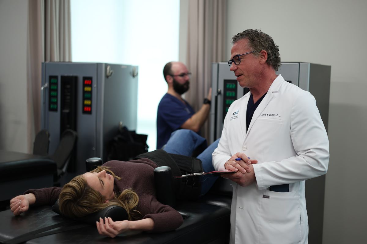
Dr. Kevin Barton has been in practice since 2003 and has run the DRX9000 since 2005. For more than twenty years he has read lumbar MRIs for a living. In February 2025 he read one of the worst he had seen, and it was his.
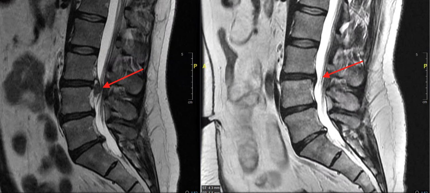
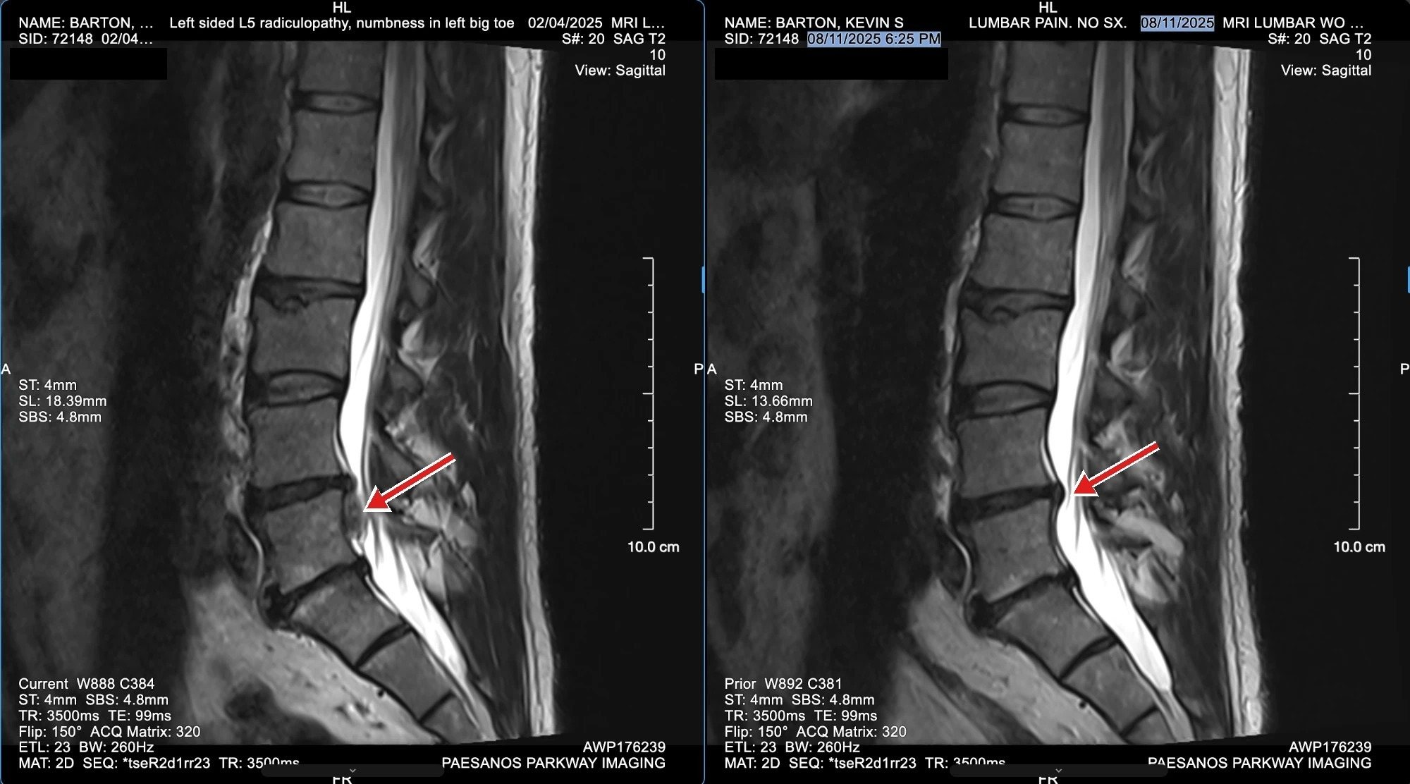
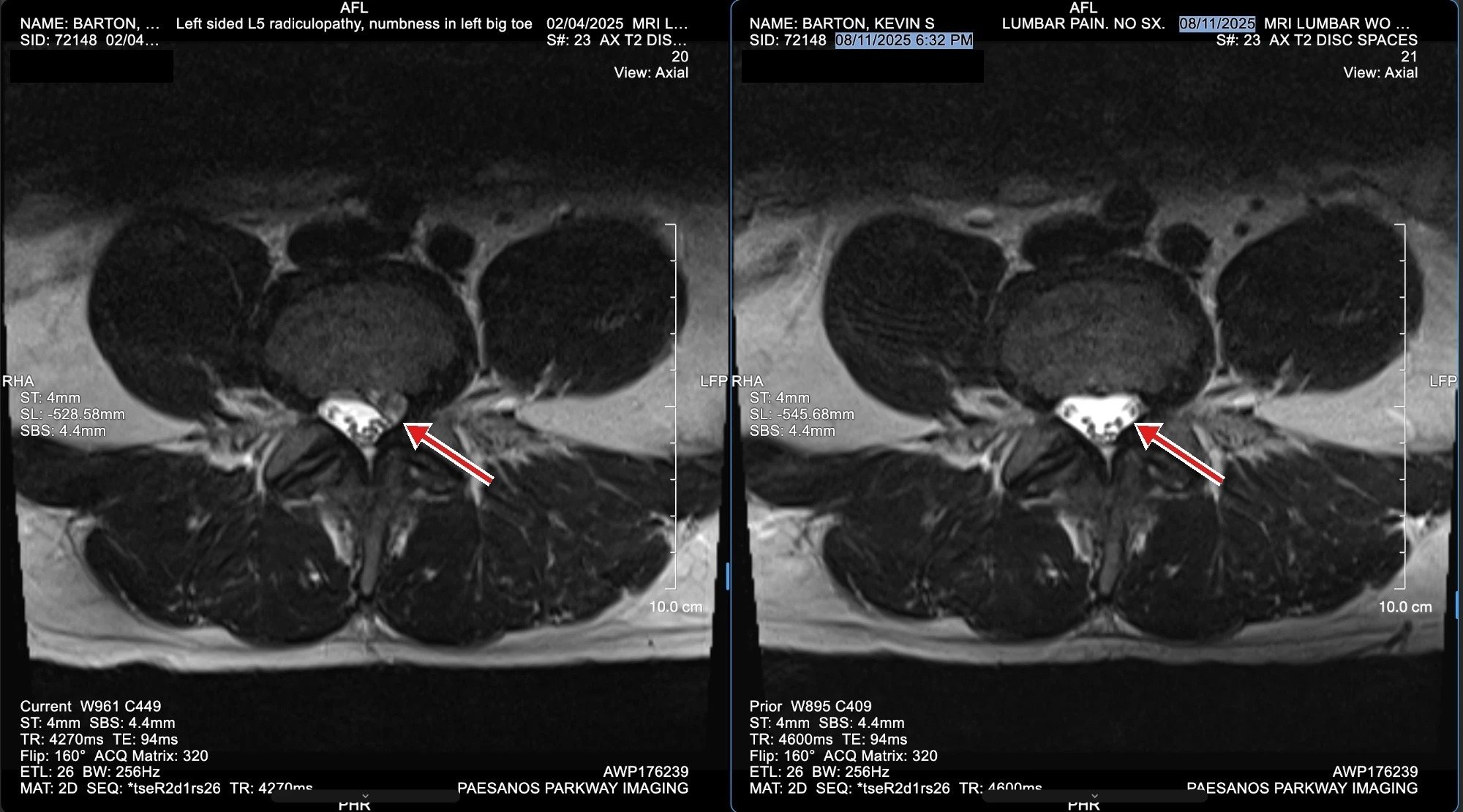
A 15 mm disc extrusion at L4-L5, completely compressing the nerve to his left leg. “My big toe was completely dead, and I had no control over it at all.” He could not walk on his heel. That combination, a large extrusion with a nerve that is already losing function, is the kind of case that gets a surgical consult.
He was a candidate for surgery. He chose the same care he offers his patients, and he filmed all of it.
Why surgery stayed off the table
Not because surgery never has a place. Because of the order things should be tried in.
Decompression is reversible. Nothing is cut, nothing is fused, nothing is implanted, and nothing about a course of treatment closes the surgical door if it is ever needed. Surgery is the opposite: it is the one option you can’t take back. When a reversible treatment has a real chance of getting the pressure off the nerve, it goes first. That is the rule he has applied to patients for two decades, and it did not change when the MRI had his name on it.
There was a second reason, and it is the one that makes the story worth telling. He knew the research on discs shrinking by themselves better than almost anyone, and he knew it wasn’t on his side. In his words:
Natural resorption probably would have taken at least a year, maybe longer. Meanwhile, the nerve would have been permanently damaged, and I would have lost use of my ankle.
A nerve that has already gone numb and weak is on a clock. Waiting is not free, and he was not willing to pay for it with an ankle.
What happened, week by week
- Week one. On the table, daily. Still unable to walk on his heel. The foot drop got worse before it got better, and that week is on camera.
- Under two weeks. Control of the big toe came back.
- Three weeks. He could perform a heel walk.
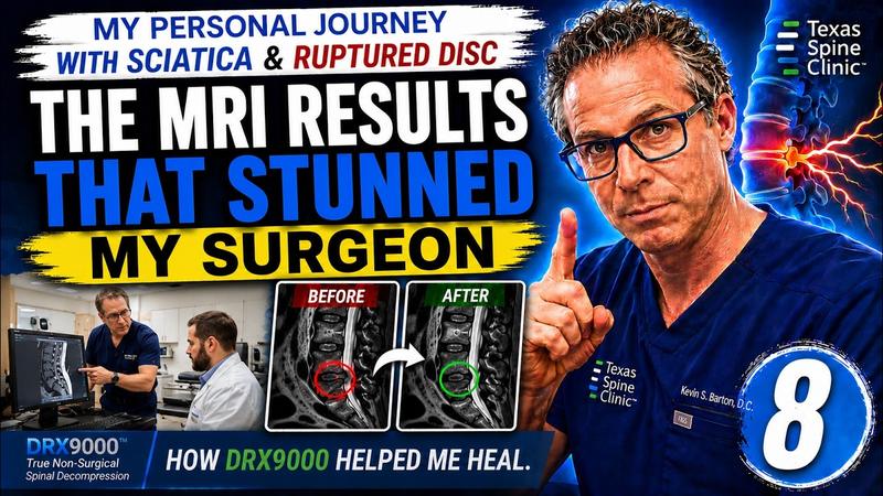
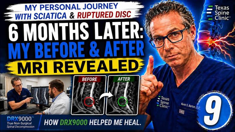
- August 2025. The follow-up MRI showed the extrusion gone. Same machine, same level, nothing left pressing on the nerve.
One case, his, proves what is possible, not what is typical. Your case is your own. It gets its own exam, its own imaging review, and its own plan. But this is why the approach here starts where it does, and why he can look a patient in the eye when he recommends it.

The whole thing, on camera
He recorded the first MRI, the exam, the sessions, the week the foot drop got worse, and the follow-up imaging at the end. Nothing edited out. Nine episodes, in the order it happened.
 Watch
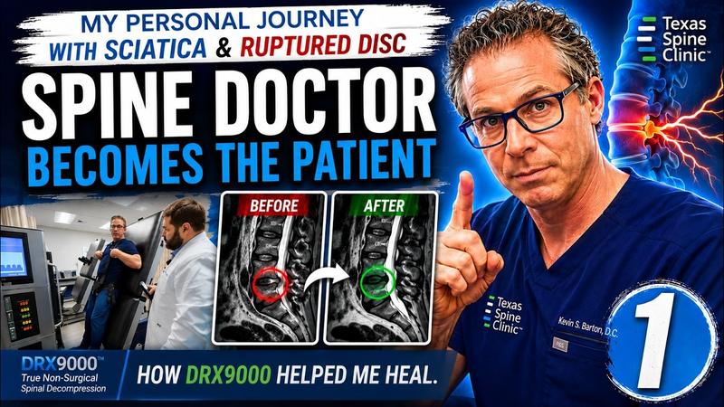
WatchEpisode 1The MRI, uneditedL4-L5 ruptured disc. His own films, on the screen.
 Watch
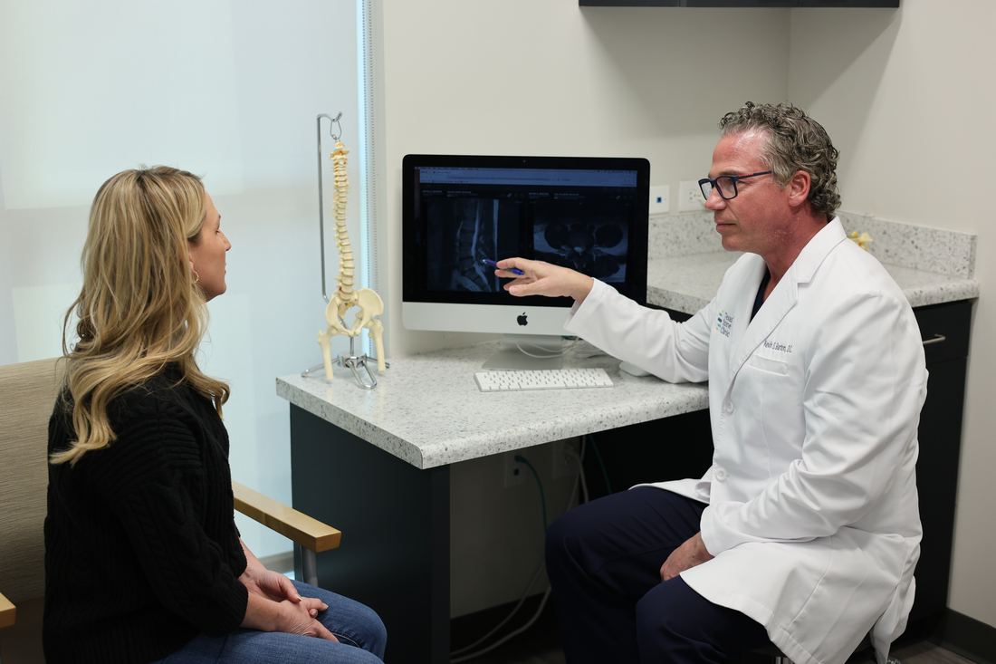
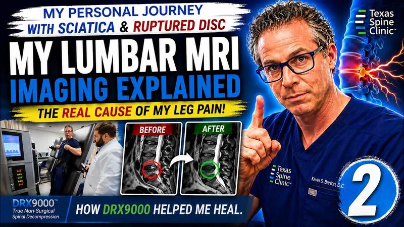
WatchEpisode 2Reading his own MRIThe test that decides whether you can avoid surgery.
 Watch
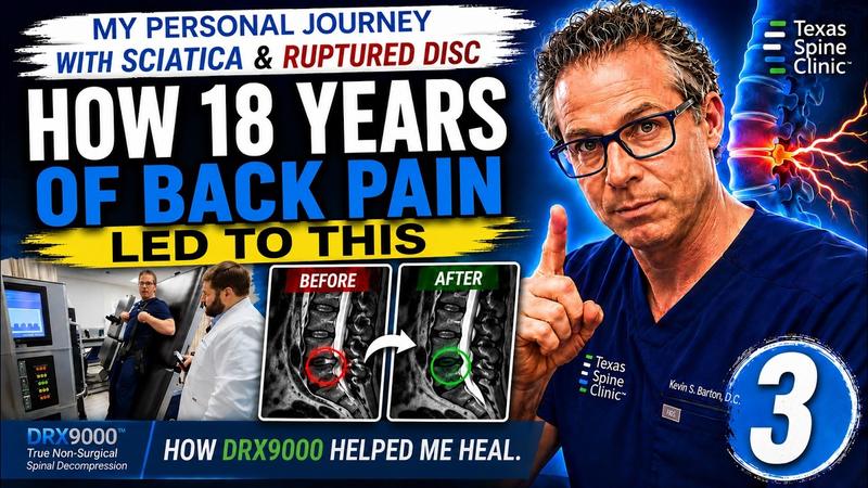
WatchEpisode 3What a proper spinal exam looks likeThe exam we run on you, run on him.
 Watch
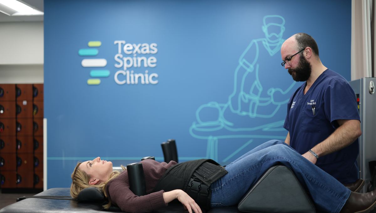
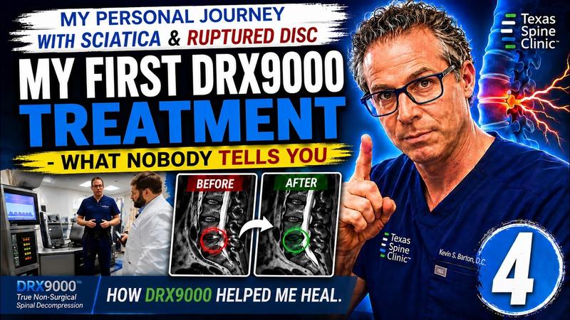
WatchEpisode 4On the table himselfTreating his own ruptured disc and sciatica on the DRX9000.
 Watch
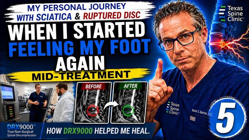
WatchEpisode 5I almost had back surgeryThe whole story in one video: what the disc did, and what he did about it.
 Watch
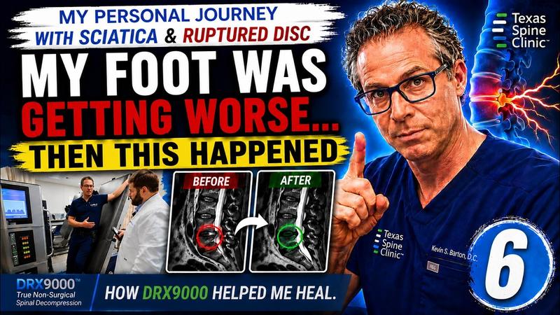
WatchEpisode 6The foot drop got worse firstThe week it got worse before it got better, on camera.
 Watch
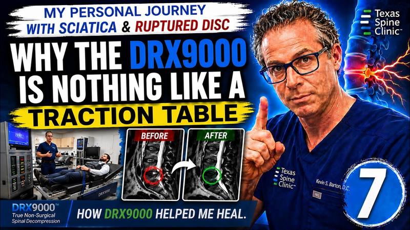
WatchEpisode 7Why this isn't tractionThree reasons the DRX9000 does something a traction table can't.
 Watch
WatchEpisode 8The MRI doesn't lieThe same disc, before and after the program.
 Watch
WatchEpisode 9Does it really work?His DRX9000 experience, start to finish.
The full playlist is on our YouTube channel: Spine Doctor’s Personal Journey With Sciatica and a Ruptured Disc.
If your MRI looks like his
A large extrusion, leg pain, a foot or a grip that is getting weaker: that is not a case to watch for a year. It is a case to examine this week. The $49 DRX9000 consultation is the same exam he ran on himself, and at the second visit your MRI goes on the same screen. He will tell you what he sees, and whether you are a candidate, because he has been on both sides of that conversation.
Book the $49 DRX9000 consultation
This article is educational and is not a substitute for an examination and diagnosis. Individual results vary and no outcome is guaranteed.